Demo Topic: Diagnostic Pacing
The concept of electrophysiology was first recognised in the 1960’s, followed by diagnostic simulation in the 1970’s and finally RFA in the 1980’s (Boyle, 2002). This section explains the main protocols and their uses: to induce, to measure, or to terminate a tachycardia.
From this point forward it is assumed that all students are familiar with the common pacing terminology explained in Pacing 1 – Fundamentals (such as “S1”, burst, BPM conversion to milliseconds, etc).
1. Protocols for EP Measurements
Train pacing (or "S1 trains")
This type of pacing is considered the simplest pacing protocol, it usually consists in delivering several pulses at a fixed rate during certain amount of time. It is normally used to test the automaticity of a node or focus. In most cases, this is achieved pacing at an slightly higher rate than the tested node or focus, then stopped and observing the later response (jumps, pauses, possible tachycardias, etc.).
The most extended use is to measure the Sinus Node Recovery Time (SNRT) at the beginning of a normal EP study. Generally, we will pace from the atrium close to the node for 30-40 seconds repeatedly (with cycle lengths of between 700ms and 300ms). After every, pacing cycle we will measure the time that the sinus node takes until recovers its normal activity.
Used for:
- Sinus Node Recovery Times
- Arrhythmia induction
- Termination of arrhythmia
- Entrainment of arrhythmias

Decremental pacing for Wenckebach periodicity
This is a routine part of the EP study, to assess the conduction from Atrium to Ventricle or from Ventricle to Atrium.
A train of S1 pacing is delivered starting at the patient`s intrinsic cycle length, and then gradually shortening the cycle length of the pacing stimulus (decrementing) every approx. 5 beats. For each stimulus delivered into the atrium, a resultant beat should occur in the ventricle (likewise from V to A).
Eventually the pacing will be delivered so fast that the AV node will not be able to keep up, and a degree of delay will slow the impulse (nodal delay).
Finally a beat will be dropped. The paced cycle length at which this occurs is termed the AV or VA Wenckebach respectively. It is important to check that if a beat appears not to conduct to the ventricle, that the atrium was in fact captured.
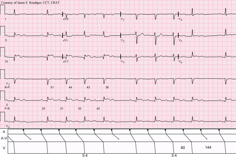
(Above) Sinus rhythm with acute inferior infarction complicated by Type I A-V block manifest in the form of 5:4 Wenckebach periods; R-P/P-R reciprocity. [Pic1]
If the terminology ‘Incremental’ and ‘Decremental’ is still unclear:

Refractory Periods Measurements (AV Node)
Extrastimulus testing in the AV Node is another routine component of the EP study, another aliases for this part of the study are: Single extras, AV Nodal Curve or AV Nodal Refractory periods.
An extra-stimuli with a conditioning train for induction of re-entrant tachycardias and refractory measurement of the AV Node.
Method:
The purpose of the 8 fixed conditioning beats is to establish reproducible cardiac conduction velocities, so the isolated effect of the varying S2 can be observed. The AV node will cause delay in the extra stimulus at shorter coupling intervals, and eventually block an early S2. When this occurs, the S1-S2 interval is termed the effective refractory period of the AV node (AVERP). Before block occurs, if enough delay is induced, the retrograde limb of a tachycardia may recover, and a re-entrant arrhythmia induced (AVNRT or AVRT).
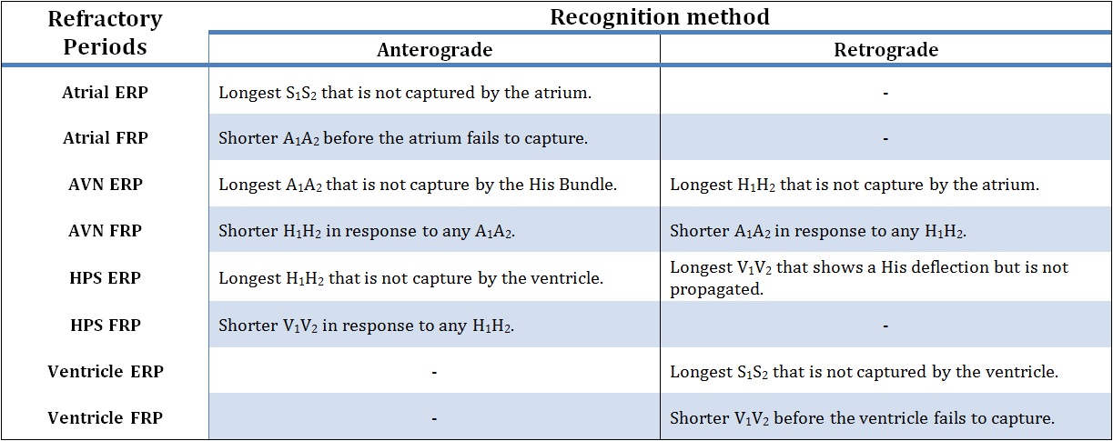
Above, possible definitions of Refractory Periods.
The extrastimulus is decreased routinely in some centers to below AVNERP until the extrastimulus fails to capture the atrium, or the Atrial ERP. This can be useful in some cases, as it may indicate myocardial conduction delay from intrinsic disease or the effect of drugs such as amiodarone or flecainide.
References:[Pic1] By Jer5150 (Own work) [CC-BY-SA-3.0 (http://creativecommons.org/licenses/by-sa/3.0)], via Wikimedia Commons.